
ACL/Tibial Plateau Leveling Osteotomy (TPLO)
Description
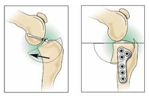
The anterior cruciate ligament (cranial cruciate ligament) is a knee ligament that may be torn or ruptured during sudden stopping or twisting movements. The resulting ligament instability causes lameness, pain, and ultimately leads to degenerative arthritis in the knee. The tibial plateau leveling osteotomy (TPLO) procedure stabilizes the knee using biomechanical principles that alter the tibial plateau slope. Rather than trying to replace the ligament, the TPLO procedure alters the angle of the knee in a way that allows the patient to function comfortably without a ligament.
Indications
There are different types of surgical procedures used to repair ruptured anterior cruciate ligament injuries. Most orthopedic surgeons prefer the tibial plateau leveling osteotomy procedure for large breed or active dogs. Regardless of the size of the patient, the conformation (shape) of the tibia should also be taken into account when choosing a stabilization procedure. Tibial plateau leveling osteotomy is indicated in any patient with a high tibial slope.
Postoperative Care
Pain medication is generally only required for the first 10 to 14 days following surgery. Give pain medication only as prescribed and do not give human drugs without first consulting with a veterinarian.
The bandage on your pet’s leg is a soft, padded bandage that controls swelling, protects the incision site, and provides some support in the early postoperative time. It is not designed to allow running or jumping. The bandage should be checked and/or changed as soon as possible if any of the following are noticed:
- Swelling of the toes occurs
- Bandage becomes wet or soiled
- Bandage has slipped
- Your pet is chewing at the bandage
If your pet has a tendency to chew, then he/she may need an Elizabethan collar designed to prevent chewing. Bandage removal is usually advised three to five days after surgery, but in some cases it may be left on until the time of suture removal.
Strict confinement should be enforced until follow-up radiographs reveal bony union at the osteotomy site. Exercise should consist of short walks outdoors only for elimination purposes on a leash. Otherwise, your pet should be kept in a pen or small room at all times located away from other pets and without furniture that he or she could jump up on. After radiographs reveal adequate healing, swimming and/or gradually increasing the leash walk distance is recommended over the next 6 weeks before normal activity is resumed.
Please schedule an appointment for suture removal 7 to 10 days after surgery. In addition, please contact us immediately if:
- Bandage problems are noted
- Any unusual swelling is noted
- Discharge of the incision site exists
- Any increase in lameness is noted after pet was starting to improve
- Limping is noticed on any of the other legs
A follow-up radiographic examination is advised 6 weeks after surgery to assess bone healing. Further follow-up may be advised depending on the rate of bone healing.
Prognosis
The prognosis for recovery to normal or near-normal function in the limb is generally excellent following a TPLO. Pre-existing arthritis may limit the full recovery of some patients, but most can regain an active, pain-free life. We have had numerous patients return to work as hunters, field trial competitors, police dogs, show dogs, and other athletic endeavors following the TPLO procedure. Potential complications following TPLO include implant failure or fracture, if exercise restriction is not enforced, and infection. The plate used to stabilize the tibia is usually left in place permanently unless a chronic infection develops. Once the bone has healed, the plate can be safely and simply removed, with no loss of stabilization of the knee, if needed to help treat infection.
Your pet’s recovery and well-being are our primary concerns, so please do not hesitate to call and speak with a surgical technician or surgeon if there are any questions regarding your pet’s recovery.
Figure 1: Voss, J. Treatment Options for Cranial Cruciate Ligament Injury/Disease of the Dog Knee (p. 2). Colorado State University. Retrieved from http://csu-cvmbs.colostate.edu/Documents/orthopaedics-cruciate-ligament.pdf.